


In the case of thoracic osteochondrosis, the limbs associated with the spinal segments, which are located at the level of the affected thoracic region and below, often suffer. Violation of the normal activity of the spine causes the arms, legs and torso to be completely immobilizedThe pelvic organs, respiratory muscles and internal organs become dysfunctional.
Osteochondrosis is a degenerative-dystrophic disease of the spine, based on changes in the intervertebral discs with involvement in the pathological process of neighboring vertebrae and intervertebral joints along with the entire ligamentous apparatus.
Features of the anatomy of the spine
The mobility and stability, elasticity and elasticity of the spinal column largely depend on the intervertebral disc, which is one of the types of cartilaginous connections between bones and provides a strong bond between neighboring vertebral bodies. Total of the intervertebral discsThe length is one quarter of the length of the spinal column.
The most important function of the disc is to reduce the vertical load on the vertebrae. The disc consists of three parts:
- hyaline plates (adjacent to the vertebrae);
- nucleus pulposus (fills the gap between the plates);
- Fibrous ring (encircles the nucleus from the outside).
The nucleus consists of cartilage cells, tightly interconnected collagen fibers and chondrins (proteoglycans). The anterior surface of the disc is covered by the anterior longitudinal ligament, which is tightly attached to the vertebrae and flips freely over the discThe posterior longitudinal ligament is firmly attached to the surface of the disc and forms the anterior wall of the spinal canal. The intervertebral disc does not have its own blood supply, so it feeds on substances that proliferate from the vertebral bodiescome by.
The distribution of vertical load in the spinal column is due to the elastic properties of the disc. As a result of pressure, the nucleus pulposus expands, and the pressure is redistributed in the annulus fibrosus and hyaline plates. During movement, the core moves in the opposite direction: when flexedis carried out - on the convex side, when unbalanced - in the anterior. When the spine moves, muscles, ligaments and discs are involved in the work. Therefore, a violation in one link leads to a violation in the entire kinetic chainIs.
Causes and mechanisms of development of the disease
In the development of osteochondrosis, a special role is played by mechanical effects on the spine. Under the influence of adverse static and dynamic loads, the nucleus pulposus gradually loses its elastic properties (as a result of depolymerization of polysaccharides), forms protrusions and sequesters.
The process of disc degeneration is influenced by a genetic predisposition, which causes changes in the neuromuscular apparatus of the back, a change in the composition of glycosamines and a violation of the distribution of collagen fibers in the discs. Genetic factors paramount in the occurrence of thoracic osteochondrosis. which is subject to increased functional activity.
Risk factors for developing degenerative changes in the spine include structural features of the discs, which are imperfections in development. One of these characteristics is the nutritional characteristics of the structures. In the human body, discs consist of poorly perfused tissue. Closure occurs in early childhood. Nutrition occurs due to the diffusion of substances through the end plates.
The stimulator of nutrient entry is a weighted load that excludes stable postures and great stress. Physical inactivity is one of the major risk factors for thoracic osteochondrosis. Therefore, regular exercise is an important preventive measure.
The peculiarity of the microscopic structure - some cells - reduces the intensity of regenerative capacity and the rate of recovery of disc components. An anatomical feature is disc fragility and lack of strength in the posterior sections. Contributes to the appearance of shaped discs.
Great importance in the development of osteochondrosis is given to involuntary changes. Actively degenerative changes begin to increase after 30 years. The synthesis of the components necessary for the discs (glycosaminoglycans) continues, but their quality is deteriorating. Hydophilicity decreases. is, fibrosis increases, sclerosis appears.
Stages of degeneration of the intervertebral disc:
- prolonged asymptomatic course, degenerative changes in intrauterine components, displacement of nuclei inside the disc;
- pronounced radicular symptoms of thoracic osteochondrosis, compression of the spinal cord, protrusion (protrusion, 1 degree) of the nucleus pulposus;
- disc rupture with hernial protrusion (hernia, 2nd degree);
- Degenerative changes (grade 3) in extradiscus components.
Pathological protrusion compresses nerve roots, blood vessels or spinal cord at different levels (cervical, thoracic, lumbar), which determines the clinical picture.
Restriction of mobility in the thoracic spine, which is due to the presence of cysts, contributes to minimal trauma to the intervertebral discs, and therefore osteochondrosis. Physiological thoracic kyphosis The weight of the upper half of the body in the lateral and anterior sections of the vertebraeContributes to redistribution. Therefore, intervertebral hernias and osteophytes are formed on the anterior and lateral surfaces of the spinal column. Posterior osteophytes and hernias are extremely rare.
Osteochondrosis contributes to the contraction of the intervertebral foramina and the compression of the roots of the spinal cord and sympathetic fibers. Sympathetic fibers originate in the gray matter of the spinal cord, then collect in nodes, sending them to all internal organs. This leads to the fact that thoracic osteochondrosis, in addition to specific neurological disorders, leads to dysfunction of internal organs (vegetative, vasomotor, trophic) and imitation of somatic diseases. This feature of osteochondrosis of the thoracic discs leads to the diagnosis of correct treatmentand explains the difficulties in determining.
Symptoms of Thoracic Osteochondrosis
Thoracic osteochondrosis is more typical for people with a sedentary lifestyle. At the same time, the load applied to the spine does not have a stimulating effect, which contributes to a disruption in the recovery of discs. Prolonged work at the computer, staggering, etc. Diseases develop in people. Such people need to do medical practice independently.
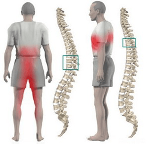
Most often, chest osteochondrosis is manifested by dull pain, less often pain and burning. The pain is localized between the shoulder blades. The patient is disturbed by a feeling of compression of the chest. When feeling the spinous processes of the thoracic vertebrae, localPain is detected, which increases with axial load on the spine, deep inspiration and twisting of the body.
Many patients have severe pain in the scapula and lower chest (posterior costal syndrome). This symptomatology develops as a result of displacement of the lower ribs. The pain becomes very sharp when turning the torso. More often, the pain syndrome suddenly disappears. Is.
Often the pain in the chest becomes girdle, corresponds to the course of the intercostal nerve. Sensitivity is disturbed in the zone of innervation of the corresponding nerve endings, paresthesia appears, and often there is a decrease in superficial and deep sensitivity. Of the abdominal press. Possible violations of function, changes in the knee and calcaneal tendon reflexes.
Violation of the function of internal organs occurs when a nerve root is compressed at levels 1 to 12 thorax. The thoracic region houses the structures responsible for innervation of the lungs, heart, intestines, liver, pancreas, and kidneys. Therefore, There are no symptoms for thoracic osteochondrosis alone.
The disease is manifested by symptoms characteristic of another pathology:
- shortness of breath;
- intense night pain;
- "heart", anginal pain;
- pain in the mammary glands;
- pain in the right or left hypochondrium (symptoms of cholecystitis and pancreatitis);
- pain in the throat and esophagus;
- pain in the epigastrium, abdomen (symptoms of gastritis, enteritis and colitis);
- sexual dysfunction.
diagnosis
The greatest value in the diagnosis of thoracic osteochondrosis is chest X-ray examination. The picture shows a decrease in the height of the intervertebral discs, sclerosis of the end plates, the formation of osteophytes.
Computed tomography allows you to determine the position of the vertebrae, the joints of the spinal column, the shape of the spinal canal, the location of the hernial protrusion and its size.
When making a differential diagnosis, it is necessary to carefully collect the anamnesis and compare all clinical signs of thoracic osteochondrosis with symptoms of other diseases. For example: pain in the heart with osteochondrosis is not stopped by nitroglycerin, epigastric pain after meals. Not associated with intake, not seasonal, all symptoms appear mainly in the evening and completely disappear after a night's rest.
How to treat thoracic osteochondrosis?
Treatment of osteochondrosis of the thoracic spine in almost all cases is conservative. The indication for therapy is the predominance of visceral syndrome with neurological disorders. The main orthopedic treatment should be adequate traction of the spine:
- active vertical traction under water;
- Passive horizontal traction in an inclined bed using Gleason loops in case of damage at the level of 1-4 thoracic vertebrae, by axial straps in case of damage at the level of 4-12 thoracic vertebrae.
Drug treatment consists in carrying out paravertebral blockade with novocaine solution. With exacerbation of the disease, analgesics and sedatives are used. With an unpredictable pain syndrome, the need to use ointments with analgesics and anti-inflammatory drugs at home. Is allowed.
After the elimination of acute events, massage of the muscles of the back and lower extremities is used. Manual therapy is indicated for 1-3 degrees of osteochondrosis in case of development of functional blockages. It consists of a gentle on the back muscles. And various options are included for the rough effect.
Therapeutic exercise allows you to load all parts of the spine in one dose, which stimulates recovery processes. An important condition for exercise therapy for osteochondrosis is to exclude vertical loads.
Physiotherapy: UHF treatment, ultrasound, inductothermy, radon and pine-coniferous salt baths. In the spa stage, underwater traction and hydromassage are actively used.
Surgical treatment is rarely used. The indication for surgical intervention is compression of the spinal cord by fragments of a prolapsed disc.